
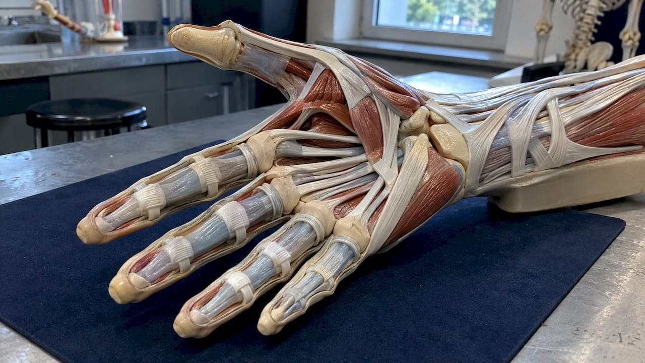
The tendons in your hand are strong, rope-like cords that connect muscles in your forearm to the bones of your fingers and thumb. Flexor tendons run along the palm side and curl the fingers. Extensor tendons run along the back of the hand and straighten them. Both groups slide inside protective sheaths and are held close to the bone by a system of pulleys. This guide walks through each part in order, shows you what a hand tendon diagram is actually depicting, and explains what happens when a tendon is injured.
TL;DR
- Hand tendons are collagen-rich cords that attach forearm muscles to the fingers and thumb, so most finger power starts in the forearm, not the hand.
- Flexor tendons bend the digits from the palm side. Extensor tendons straighten them from the back of the hand.
- Sheaths let tendons glide, and pulleys keep them against the bone so the fingers can close smoothly.
- A cut, a sudden loss of finger bending or straightening, or a finger that will not move on its own needs prompt assessment by a doctor or hand specialist.
- Healing is slow and happens in overlapping phases, and guided hand therapy is a major part of the outcome.
Table of Contents
- What a tendon is and why the hand depends on them
- Flexor tendons: the palm side
- Extensor tendons: the back of the hand
- Sheaths and pulleys: what lets tendons glide
- Reading a hand tendon diagram
- Common hand tendon injuries and warning signs
- How hand tendon injuries heal and what recovery looks like
- Our perspective
- Explore the hand in 3D
- Tendons elsewhere in the body
- FAQ
- Sources
What a tendon is and why the hand depends on them
A tendon is a band of connective tissue that transmits the pull of a muscle to a bone. Under the microscope, tendons consist of long collagen fibres arranged in bundles, with elastic fibres and small vessels running between them. That structure is why a tendon is strong under tension but has limited blood supply and heals slowly.
The hand is unusual because most of the muscle power for gripping and finger movement is not in the hand at all. The flexor and extensor tendons belong to what clinicians call the extrinsic system: the muscles start proximal to the hand, often in the forearm, and send long tendons across the wrist. This keeps the fingers slim and nimble, but it also means the tendons are exposed to injury along their length, especially on the palm side and over the back of the knuckles where there is little soft tissue covering them.
A second group, the intrinsic muscles, sits inside the hand. The lumbricals, for example, arise from the deep flexor tendons in the palm and help coordinate finger movement. They are worth knowing about because they link the flexor and extensor systems.

Flexor tendons: the palm side
Flexor tendons bend the fingers and thumb. In the fingers there are two layers:
- Flexor digitorum superficialis (FDS): attaches to the middle bone of each finger and bends the middle joint (PIP joint).
- Flexor digitorum profundus (FDP): lies deeper, passes through a split in the FDS tendon and attaches to the tip bone, bending the fingertip joint (DIP joint).
- Flexor pollicis longus (FPL): the long flexor of the thumb, which bends the thumb tip.
The FDS and FDP tendons pass through the carpal tunnel at the wrist and then glide within the finger pulley systems. The median nerve shares the carpal tunnel with them, which is why swelling there can affect both tendon glide and sensation in the thumb, index and middle fingers.
Because these tendons run so close to the surface of the palm and fingers, flexor tendon injuries tend to come from cuts. Flexor tendons usually need surgical repair when they are fully divided.
Extensor tendons: the back of the hand
Extensor tendons straighten the fingers and thumb and extend the wrist. Their muscles sit on the back of the forearm. The tendons enter the hand through six dorsal compartments at the wrist, held in place by a band called the extensor retinaculum, much like a watchband keeps a cable close to the wrist.
Key extensor tendons include:
- Extensor digitorum communis (EDC): straightens the four fingers, one tendon to each finger.
- Extensor indicis and extensor digiti minimi: give the index and little fingers some independent extension, which is why you can point or extend the little finger on its own.
- Thumb extensors and abductors: a group that includes the tendons along the thumb side of the wrist, which is the area involved in De Quervain's tenosynovitis.
On the back of the hand, the EDC tendons are linked by small bridges called juncturae tendinum. That is why, when one finger is held down, its neighbours cannot always lift fully. Over the finger itself, the tendon spreads into a hood that wraps around the knuckle joint and, through central and lateral bands, ends on the tip bone of the finger.
Extensor tendons are thinner and flatter than flexor tendons. Most tendon injuries need surgery, but some blunt injuries where the extensor tendon pulls off near the fingertip may be treated without surgery, usually with splinting. A clinician has to decide which applies.
Sheaths and pulleys: what lets tendons glide
A tendon that simply ran straight from wrist to fingertip would bowstring away from the bone every time you made a fist. Two structures prevent that.
Tendon sheaths
A thin covering called the peritendineum, or tendon sheath, provides the slippage tendons need. In the hand, flexor tendons are enclosed in sheaths that lubricate them and reduce friction as they travel beneath the transverse carpal ligament. When a sheath becomes inflamed, the condition is called tenosynovitis.
Finger pulleys
The fibrous tunnel over each finger includes four to five annular pulleys (A1 to A5) and three weaker cruciate pulleys (C1 to C3) that hold the flexor tendons close to the bone. Annular pulleys are the strong, ring-like bands. Cruciate pulleys are thinner, criss-cross bands that fold as the finger bends, allowing the finger to close.

The A1 pulley sits at the base of the finger in the palm. It is the area involved in trigger finger, where the tendon catches as it passes through a thickened pulley. The A2 and A4 pulleys, over the bones of the finger, are generally the most important for preventing bowstringing.
Reading a hand tendon diagram
A good hand tendon diagram usually shows several layers, and it helps to know what to look for:
| What you see | What it represents |
|---|---|
| Long cords on the palm side, passing through the wrist | Flexor tendons (FDS, FDP, FPL) in the carpal tunnel |
| Cords on the back of the hand fanning to each finger | Extensor tendons, often joined by juncturae |
| Ring-like bands over the finger bones | Annular pulleys (A1 to A5) |
| Thinner criss-cross bands between rings | Cruciate pulleys (C1 to C3) |
| Sleeve around the tendon | Synovial sheath |
| Small muscles beside the flexor tendons in the palm | Lumbricals |
| Band across the back of the wrist | Extensor retinaculum |
Most diagrams are flat, which hides the key fact: the FDP tendon passes through the FDS tendon. A 3D view makes the depth much easier to understand. Our wrist and hand anatomy guide covers the bones, nerves and tendons together, so you can see how they relate rather than learning each in isolation.
Common hand tendon injuries and warning signs
Tendon injuries are among the most common hand injuries. They tend to fall into a few patterns:
- Cuts and lacerations: a wound over the palm or finger can divide a flexor tendon, often with no pain proportional to the damage. The finger may simply rest straighter or more curled than its neighbours.
- Avulsion injuries: a tendon pulls away from its bone attachment, often during sport when a finger is caught.
- Overuse and tendinopathy: irritation from repeated gripping, typing or lifting, such as at the thumb side of the wrist.
- Trigger finger: catching or locking of a finger as the tendon passes the A1 pulley.
- Rupture: sudden loss of movement, sometimes with a snap, in a finger or thumb.
When to get assessed quickly
See a doctor, urgent care or emergency department promptly if you have:
- A cut on the palm, finger or wrist, particularly if you cannot fully bend or straighten a finger.
- A finger that sits in an unusual position or drops at the tip after an injury.
- Numbness, tingling or colour change in the fingers along with the injury.
- Sudden loss of grip strength or a finger that will not move when you try.
A finger that still moves a little can still have a partly or completely divided tendon, so movement alone does not rule out damage. If you are unsure, have it checked. Repair outcomes depend on getting the right treatment, and a hand surgeon or hand therapist is the right professional for tendon problems.
How hand tendon injuries heal and what recovery looks like
Tendons heal in three overlapping phases: inflammation in roughly the first 48 hours, proliferation from about 7 to 21 days, and remodelling that begins months after injury and can continue for 12 months or more. The practical meaning is that a tendon can feel better long before it has regained its strength.
Two features of tendon biology shape rehabilitation:
- Tendons heal partly from within, supported by tendon movement and cells called tenocytes, and partly from outside, through a more distinct inflammatory response that can cause scarring and adhesions.
- Repaired tissue is biomechanically weaker than the original and often scar-like, which is why protection and gradual loading both matter.
After flexor tendon surgery, early passive movement is important, and physiotherapy and occupational therapy are essential to the result. In practice, a hand therapist usually makes a splint and teaches a protocol of controlled movement. Follow that protocol exactly. Doing too much too soon risks rupture, and doing too little risks stiffness.
What a typical rehab pathway looks like
Exact timelines are set by your surgeon and therapist, because they depend on which tendon was injured, how it was repaired and your own health. In general the stages are:
- Protection: splint and swelling control, with the gentle movements you have been shown.
- Controlled motion: guided movement to keep the tendon gliding and limit scarring.
- Active strengthening: gradual loading once the repair can tolerate it.
- Return to function: grip, work and sport demands built back step by step.
If your problem is irritation rather than a tear, such as pain along the thumb side of the wrist, our De Quervain's tenosynovitis recovery plan and wrist sprain recovery plan lay out the stages and exercises. For the thumb's inner ligament, see the thumb ulnar collateral ligament plan.
Our perspective
Most people meet hand tendon anatomy for the first time after an injury, and the first diagram they find is a flat drawing with a dozen unfamiliar labels. We think that is the wrong way round.
The tendons make sense once you see them as part of a connected system: a muscle in the forearm, a cord crossing the wrist, a sheath, a pulley and a bone. When you know which structure is involved, the symptoms, the treatment conversation and the exercises all become easier to follow.
That is why we built InjuryAtlas around anatomy first. You can start from a symptom, a structure or a training goal and follow connected guides from there, in plain language, without needing a medical dictionary. Our guides are meant to help you ask better questions of your doctor or hand therapist, not to replace them.
Josh
Explore the hand in 3D
If you want to see how the tendons, bones and nerves of the wrist and hand fit together, start with our wrist and hand anatomy guide. It links to connected guides for specific injuries and recovery plans.

If you have pain but do not yet know the cause, you can also find an injury by symptom or location, or browse the exercise library by region and difficulty.
Tendons elsewhere in the body
The same principles of structure apply beyond the hand: collagen fibres, limited blood supply and slow remodelling. If you are curious how the pattern repeats, compare with the patellar tendon at the knee and the Achilles tendon at the ankle. Higher up the arm, the distal biceps tendon recovery plan covers a tendon that attaches near the elbow.
FAQ
How many tendons are in the hand?
There is no single tidy number, because it depends on whether you count each finger's separate flexor and extensor tendons, the thumb tendons and the wrist tendons. What matters is the pattern: flexors on the palm side, extensors on the back, and both starting from muscles in the forearm.
What is the difference between a tendon and a ligament in the hand?
A tendon connects muscle to bone and transmits the force that moves a joint. A ligament connects bone to bone and stabilizes the joint. The thumb ulnar collateral ligament, for example, steadies the thumb's base joint, while the flexor tendons move it.
Can a finger still move if a tendon is cut?
Yes, sometimes. A partly cut tendon, or a finger moved by a neighbouring tendon, can still show some movement. Any cut on the palm or finger with weakness or an odd finger position should be assessed promptly by a doctor or hand specialist.
How long does hand tendon recovery take?
It varies with the injury and treatment. Biologically, tendon healing continues through remodelling for 12 months or more, even though daily use usually returns earlier. Your surgeon and hand therapist will give a timeline for your specific tendon.
Sources
- Tendon injuries of the hand (PMC)
- Tendon: Principles of Healing and Repair (PMC)
- Tendinopathies, Handthology by ASSH
- Extensor muscle overview, ScienceDirect Topics
- Flexor Tendons: Anatomy and Surgical Approaches