
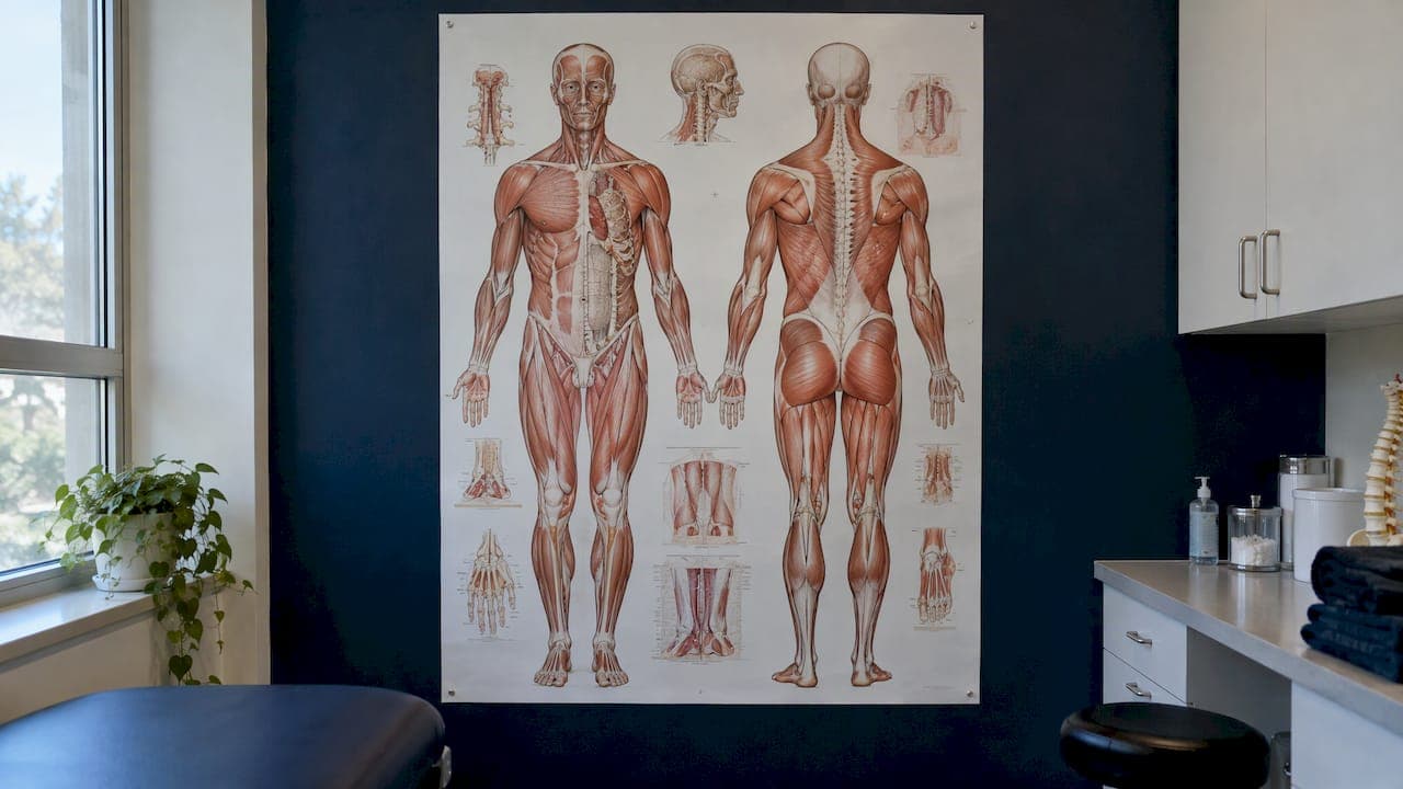
A human anatomy chart is a labelled map of the body. It shows where bones, muscles, nerves, vessels and organs sit, usually from the front and the back, and it is most useful when you read it by region and then connect each region to the systems running through it. If you are trying to understand a sore shoulder, a stiff lower back or a knee that aches on stairs, the chart gives you the vocabulary and the location. It does not give you a diagnosis, and this guide explains where that line sits.
Below you will find how to read front and back views, the main body regions and what lives in each, how a body pain chart helps and where it stops, how to describe symptoms to a clinician, and what to look for when choosing a chart.
TL;DR
- An anatomy chart shows structures in position. Front (anterior) and back (posterior) views are drawn as if the person is facing you, so the left side of the page is the person's right side.
- Reading by region (head and neck, trunk, upper limb, lower limb) is the easiest way to learn, and each region contains parts of several body systems.
- A body pain chart tells you what lies under the spot that hurts. It cannot tell you why it hurts, because pain in one place can come from several structures.
- Bring the location, start, pattern and triggers of your pain to a clinician. Some symptoms, such as numbness in the saddle area with bladder or bowel changes, need emergency care.
- For study, choose a chart that labels clearly, separates layers (bones, muscles, nerves) and links each structure to plain-language explanations.
Table of Contents
- What a human anatomy chart actually shows
- Body regions and what sits in each
- How regions relate to body systems
- Using a body pain chart without over-reading it
- Explaining your symptoms to a health professional
- When to see a professional instead of reading a chart
- Choosing an anatomy chart for study, patient education or reference
- Our perspective
- Explore the body region by region with InjuryAtlas
- FAQ
- Sources
What a human anatomy chart actually shows
Most charts present the body in the standard anatomical position: standing upright, facing forward, arms at the sides, palms forward. Everything is described relative to that pose, so a structure on the "anterior" surface is toward the front, and "posterior" is toward the back. "Superior" means toward the head, "inferior" toward the feet, "medial" toward the midline and "lateral" away from it.

One detail trips up many readers: left and right are the person's own. On a front view, the figure's right shoulder appears on the left side of the page. If you are matching a chart to your own body in a mirror, remember that the mirror flips this again, which is a common source of confusion when people try to find the "right" side.
Front and back views
The front view typically shows the face, neck, chest, abdomen, front of the shoulders, the biceps side of the arm, the front of the thighs and the shins. The back view shows the skull base, the spine, shoulder blades, the back of the arms, buttocks, hamstrings and calves. If your pain is at the back of the body, a front-only chart will not help, so look for a set that offers both.
Layers matter more than detail
A chart that shows only skin-level landmarks is of limited use for pain. Good charts separate layers: surface muscles, deeper muscles, the skeleton, nerves and blood vessels. A pain on the outer hip, for example, could involve the bone, a bursa, a tendon or a muscle, and you can only reason about it when you can see each layer on its own.
Body regions and what sits in each
Regional anatomy divides the body into zones and studies everything inside each zone together. It is the approach that matches how pain is felt, because you feel pain in a place, not in a system.
| Region | Main structures | Common reasons people look it up |
|---|---|---|
| Head and neck | Skull, jaw joint, cervical spine, neck muscles, major nerves and vessels | Neck stiffness, jaw pain, headaches that start in the neck |
| Upper back and chest | Thoracic spine, ribs, shoulder blades, heart and lungs behind the rib cage | Pain between the shoulder blades, rib pain on breathing |
| Shoulder, arm and elbow | Collarbone, shoulder joint, rotator cuff, biceps, triceps, elbow tendons | Shoulder pain at night, tennis or golfer's elbow |
| Wrist and hand | Carpal bones, tendons, median and ulnar nerves | Thumb-side wrist pain, tingling fingers |
| Lower back and abdomen | Lumbar spine, back muscles, abdominal wall, digestive and urinary organs | Lower back pain, flank or abdominal pain |
| Hip and groin | Hip joint, pelvis, hip flexors, adductors | Groin pain, hip pinching on sitting |
| Knee | Femur, tibia, patella, ACL, PCL, MCL, LCL, menisci | Pain after a twist, swelling, instability |
| Lower leg, ankle and foot | Tibia, fibula, ankle joint, Achilles and other tendons, foot bones | Shin pain, ankle sprain, big toe sprain |
The spine is a good example of regional naming. The vertebral column is divided into five regions: cervical, thoracic, lumbar, sacral and coccygeal. The same source describes how the abdomen is mapped into four quadrants or nine regions so that tenderness can be located precisely. If you want to see two of these spinal regions in detail, our guides to the thoracic spine from T1 to T12 and the lumbar spine show the bones, discs, joints and muscles in each.
Abdominal quadrants and why they exist
The four-quadrant system divides the abdomen with one vertical and one horizontal line through the navel: upper right, upper left, lower right and lower left. Clinicians use it as shorthand. When someone says "lower right abdominal pain," the quadrant narrows the list of organs underneath. The nine-region version adds more precision and is more common in anatomy teaching.
Regions in the limbs
Limbs are usually broken into segments: the shoulder girdle, arm, forearm, wrist and hand for the upper limb, and the hip, thigh, knee, leg, ankle and foot for the lower limb. Each segment has its own bones, muscles and nerve supply, which is why a problem at the wrist can send tingling into specific fingers. Our wrist and hand anatomy guide and hip joint overview follow this segment-by-segment logic.
How regions relate to body systems
A region is a place. A system is a network that runs through many places: the skeletal, muscular, nervous, cardiovascular, respiratory, digestive, urinary, endocrine, immune and reproductive systems, plus the skin. Many charts and 3D tools are organized by system, which is why the top search results for this topic often list a dozen of them.
Both views are valid and they complement each other. Regional teaching is common in anatomy courses, where students study regions while integrating the relevant body systems, according to one anatomy textbook chapter. In practice that means that when you look at the lower back, you are not looking at "just muscle." You are looking at vertebrae and discs (skeletal), the back muscles (muscular), the spinal nerves (nervous), the arteries supplying them (cardiovascular), and, in the front of the same region, the kidneys and bowel.
This is why pain can be confusing. The quadratus lumborum, a deep muscle of the low back, sits near the kidneys, and our quadratus lumborum guide explains how a muscle problem there can feel like a deeper, more worrying ache. A chart that shows both layers helps you see why two very different problems can feel similar.
Using a body pain chart without over-reading it
A body pain chart is an anatomy chart with a purpose: you mark where it hurts and read off the structures underneath. Done well, it narrows your questions. Done poorly, it can make you decide on a cause too early.
What it can tell you
- What lies beneath the painful spot. This gives you names to look up and use in conversation.
- What lies near it. Referred pain, where the source is not where you feel it, makes neighbouring structures relevant.
- How to describe the location precisely. "Outer side of the right knee, just below the joint line" is far more useful than "knee pain."
What it cannot tell you
Location is a clue, not a verdict. Medical references show this clearly. In the abdomen, for example, the site of tenderness can point to the problem, and tenderness at a spot called McBurney point is likely with appendicitis. Yet the same reference stresses that a provider needs to assess it. Likewise, flank pain can be a sign of a kidney problem, but other causes are possible, including muscle strain.

That pattern holds across the body. Pain at the front of the shoulder might come from the biceps tendon, the rotator cuff or the joint itself. Groin pain can arise in the hip joint, a muscle, the abdominal wall or elsewhere. Our guides on groin pain, lower back pain and shoulder pain at night each list the usual candidates so you can see the range of possibilities rather than fixing on one.
A simple way to use it
- Find the painful area on both the front and back views.
- Note every structure listed under it, layer by layer.
- Look at the neighbouring regions too, especially above and below the spot.
- Write down the names that seem to fit your symptoms, as questions rather than conclusions.
- Take those notes to a clinician.
Explaining your symptoms to a health professional
A marked-up chart is a good way to show where something hurts, especially when the words are hard to find. But the location is only one piece of the story. Clinicians commonly ask about where the pain is, when it began, whether it is constant or comes and goes, and whether it is getting worse. They also ask what you were doing, what makes it better or worse, and whether other symptoms appear alongside it.
Before an appointment, it helps to prepare a short note covering:
- Where: the exact spot, using chart terms if you know them, plus any place the pain travels to.
- When it started: a sudden injury, a gradual build-up, or after a particular activity.
- Pattern: constant, intermittent, worse at night, worse in the morning.
- Triggers: movements, positions or activities, such as bending, lifting or climbing stairs.
- What it feels like: sharp, dull, burning, aching, tingling or numb.
- Other symptoms: swelling, fever, weakness, locking or giving way.
If you are not sure which injury fits, the Find an Injury page lets you start from the area or symptom and see connected guides. It is a way to prepare better questions, not to replace an assessment.
When to see a professional instead of reading a chart
A chart is a poor tool when something serious might be happening. Seek assessment promptly for pain after significant trauma, an obvious deformity, inability to bear weight, severe swelling, fever with a hot joint, or pain that keeps worsening. Some situations are emergencies.
- Breathing difficulty or heavy bleeding. These are emergency symptoms. MedlinePlus lists them in its guidance on a broken or dislocated jaw, which also needs prompt medical attention.
- Possible cauda equina syndrome. If back pain comes with numbness around the saddle area or new bladder or bowel problems, go to emergency care. A UK National Health Service document on cauda equina syndrome says it requires immediate emergency assessment and lists these as possible symptoms.
- Chest, abdominal or flank pain that is severe or unexplained. The structures under these regions include organs, and only an examination can sort them out. In Canada, you can call 911 for emergencies or 811 for non-urgent health advice in most provinces.
If you have just injured a joint and are unsure how serious it is, a family doctor, walk-in clinic, physiotherapist or sports medicine clinician can examine it and decide whether imaging is needed. Bring your notes and any marked chart.
Choosing an anatomy chart for study, patient education or reference
The search results for this topic are dominated by printed posters, laminated wall charts and 3D explorers. They serve different needs, and the right one depends on what you are trying to do.
| Type | Best for | Strengths | Limits |
|---|---|---|---|
| Printed wall chart or poster | Classrooms, clinics, a study wall | Always visible, no screen needed | One layer per chart, fixed view, little explanation |
| Chart set (muscular, skeletal, nerves) | Studying layers in turn | Covers several systems | You switch between sheets to follow one region |
| Interactive 3D explorer | Rotating structures and seeing depth | Any angle, layers can be toggled | Often system-based, with limited injury context |
| Region-and-injury guides | Understanding a specific pain | Connects structure, symptom and recovery | Covers the regions the guide includes |
What to check before you choose
- Both front and back views. Many pain problems sit at the back of the body.
- Separate layers. Bones, muscles, nerves and vessels should be distinguishable.
- Clear, consistent labels. Look for standard anatomical terms, with plain-language names alongside.
- Left and right handled clearly. Good charts state that labels follow the person's own sides.
- A way to go further. A chart with no explanation leaves you with names but no meaning. For patient education, pair it with text that explains what each structure does and what typically goes wrong.
- Fit for the audience. A physiotherapist's reference, a student's study aid and a patient handout need different levels of detail.
For patients, a simple chart marked up with the clinician during an appointment is often more helpful than a very detailed one. For students, layered detail and repeated testing matter more. For athletes, region-specific guides that connect a structure to the injuries it commonly suffers are usually the most practical.
Our perspective
We built InjuryAtlas around a simple observation: a chart on its own gives you names, but people come to anatomy because something hurts, or because they want to train without getting hurt. Names are not enough.
That is why we treat anatomy as the centre and connect everything else to it. Every symptom, injury and exercise on the platform is tied to the specific structure involved, so you can start from a pain, a structure or a training goal and follow a connected path. We write in plain language, mix in the technical terms you will hear in clinic, and aim to keep the tone calm, because worry is rarely helpful when you are trying to understand your body.
We also think a good chart should be honest about its limits. Where you feel pain is a strong starting point, and we design our guides to help you ask better questions and describe symptoms clearly. Diagnosis belongs to a clinician who can examine you, and we encourage that conversation. Our aim is for you to walk into the appointment informed, and for physical therapists and students to have a reliable reference they can check quickly.
Josh
Explore the body region by region with InjuryAtlas
If you want more than a static diagram, InjuryAtlas lets you explore anatomy in 3D, pinpoint where it hurts and follow connected guides on symptoms, recovery and exercises for the upper body and knee. Our library includes more than 79 anatomy guides plus encyclopedia entries, all written in plain language.

Start on the InjuryAtlas home page, browse the encyclopedia, or use the whole-body symptom encyclopedia when you know the symptom but not the structure. For movement, the exercise library is organized by region and difficulty, and the knee recovery section brings the structures, injuries and rehab steps together.
FAQ
What is the difference between a human anatomy chart and a human body anatomy chart?
They describe the same thing. Both show the structures of the body in labelled form. The difference is usually scope: some charts cover the whole body at a glance, while others focus on one system, such as muscles or the skeleton, or on one region, such as the knee or the spine.
How do I read the left and right sides on an anatomy chart?
Charts follow the anatomical position and label sides from the person's point of view. On a front view, the figure's right side appears on the left of the page. Look for "R" and "L" markers, and remember that a mirror reverses the pattern again.
Can a body pain chart tell me what is wrong?
No. It shows which structures lie under and near the painful area, which narrows your questions. Several different problems can cause pain in the same place, and the cause is confirmed by a history and examination from a health professional.
What are the main body regions in anatomy?
The usual divisions are head and neck, trunk (chest, abdomen, back and pelvis), upper limbs and lower limbs. The spine is further divided into cervical, thoracic, lumbar, sacral and coccygeal regions, and the abdomen is mapped into quadrants or nine regions.
Which anatomy chart is best for patient education?
Choose one with clear labels, front and back views and separate layers, then mark it up with the clinician during the visit. Pair it with plain-language explanations of what each structure does, so the names have meaning for the patient.
Sources
- 2.4: Body Regions, Medicine LibreTexts
- Chapter 1: Introduction to the Human Body, Elsevier eLibrary
- Point tenderness, abdomen: MedlinePlus Medical Encyclopedia
- Flank pain: MedlinePlus Medical Encyclopedia
- Broken or dislocated jaw: MedlinePlus Medical Encyclopedia
- Cauda Equina Syndrome Awareness, Oxford University Hospitals NHS