
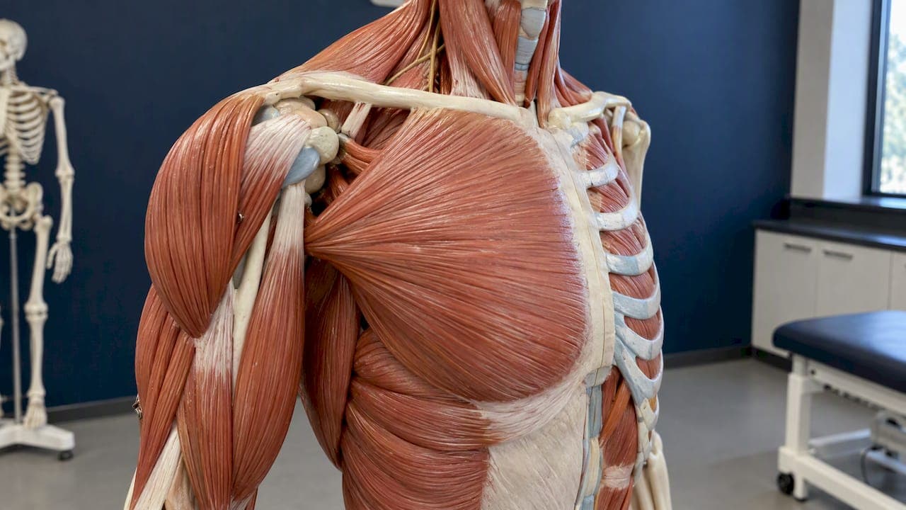
The shoulder is moved and held together by about a dozen muscles in four working groups: the four rotator cuff muscles that centre the ball in its socket, the deltoid that lifts the arm, the scapular muscles that position the shoulder blade, and the chest and back muscles (pectoralis major and latissimus dorsi) that drive big pushing and pulling movements. This guide walks through each group, shows what a good labelled diagram should include, and explains how muscle and tendon problems show up as pain.
TL;DR
- The rotator cuff (supraspinatus, infraspinatus, teres minor, subscapularis) keeps the head of the humerus centred in the shallow shoulder socket.
- The deltoid is the large outer "cap" of the shoulder and does most of the lifting of the arm away from the body.
- The pectoralis major, the main chest muscle, has two heads with separate nerve supplies and pulls the arm forward and inward.
- Scapular muscles such as the trapezius and serratus anterior set the shoulder blade so the arm has a stable base.
- Pain that follows a sudden injury, comes with weakness, or wakes you at night deserves a professional assessment.
Table of Contents
- Why the shoulder needs so many muscles
- The rotator cuff: four muscles, one job
- The deltoid: the shoulder's powerhouse
- Scapular muscles: the moving foundation
- Chest muscle anatomy: pectoralis major and minor
- Other muscles that cross the shoulder
- How the muscles work together
- When muscle or tendon problems cause shoulder pain
- Our perspective
- Explore the shoulder in 3D
- FAQ
- Sources
Why the shoulder needs so many muscles
The shoulder is the most mobile joint in the body, and that freedom comes at a cost. The upper arm bone (humerus) has a rounded head that sits against a small, shallow socket on the shoulder blade (scapula). There is very little bone holding the ball in place, so muscles and their tendons do most of the stabilizing.
A useful way to organize the anatomy is by job rather than by name:
- Centre the joint: the rotator cuff.
- Lift and move the arm: the deltoid, with help from the biceps and chest.
- Position the shoulder blade: trapezius, serratus anterior, rhomboids, levator scapulae and pectoralis minor.
- Produce large forces: pectoralis major, latissimus dorsi and teres major.
If you want to explore these structures in three dimensions and see how they overlap, our Shoulder Anatomy Encyclopedia groups every muscle, tendon and joint on one connected map.
The rotator cuff: four muscles, one job
The rotator cuff is a group of four muscles that begin on the shoulder blade and wrap around the head of the humerus. Their tendons blend together into a cuff that attaches to the top of the upper arm bone. Because of this arrangement, the cuff acts like a set of adjustable guy-wires, pulling the ball snugly into the socket while larger muscles move the arm.
| Muscle | Where it sits | Main action |
|---|---|---|
| Supraspinatus | Top of the shoulder blade, runs under the bony roof of the shoulder (acromion) | Starts lifting the arm out to the side and compresses the ball into the socket |
| Infraspinatus | Back of the shoulder blade, below its bony ridge | Rotates the arm outward |
| Teres minor | Lower outer edge of the shoulder blade, just below infraspinatus | Rotates the arm outward and helps hold the ball down in the socket |
| Subscapularis | Front surface of the shoulder blade, against the rib cage | Rotates the arm inward and resists the ball sliding forward |
What a rotator cuff muscles diagram should show
A good diagram uses at least two views, because the cuff cannot be seen well from one side. Look for:
- A back view that labels the supraspinatus above the scapular spine and the infraspinatus and teres minor below it.
- A front view (or a view with the rib cage removed) that shows the subscapularis on the underside of the shoulder blade.
- The attachment points on the humerus: the greater tubercle on the outer top of the bone and the lesser tubercle at the front.
- The tendons drawn separately from the muscle bellies, so you can see where muscle turns into tendon.
- The long head of the biceps tendon, which runs in a groove between the two tubercles and sits right beside the cuff.
What a rotator cuff tendons diagram adds
The tendon version of the diagram matters when you are trying to understand pain. It shows how the supraspinatus tendon passes through a narrow space under the acromion, and where the subacromial bursa (a small fluid-filled cushion) lies above it. That tight passage is why the supraspinatus tendon is the one most often irritated or worn. Seeing the tendons in place makes it easier to understand why raising the arm overhead can pinch or aggravate a sore cuff.

If you are rehabbing a cuff problem, our guide to the 5 best rotator cuff exercises shows how to load these four muscles gradually.
The deltoid: the shoulder's powerhouse
The deltoid is the thick, triangular muscle that gives the shoulder its rounded outline. It starts on the collarbone (clavicle), the acromion and the spine of the scapula, and it attaches to a rough spot partway down the outer humerus. It has three parts, and each does something slightly different:
- Front (anterior) fibres: lift the arm forward and rotate it inward.
- Middle (lateral) fibres: lift the arm out to the side. This is the part that does most of the work in a lateral raise.
- Back (posterior) fibres: pull the arm backward and rotate it outward.
The rotator cuff and deltoid work as a team. When you raise your arm, the deltoid pulls the humerus upward while the cuff pulls it into the socket. If the cuff is weak or torn, the deltoid can push the ball upward against the acromion, which is one reason cuff problems can feel like pinching at the top of the shoulder. You can read more about this muscle on our deltoid anatomy page.
Scapular muscles: the moving foundation
The shoulder blade is not fixed. It glides across the back of the rib cage every time you move your arm, and the muscles that control it decide whether the socket points in a useful direction. These are sometimes called the extrinsic shoulder muscles, because they start on the trunk and attach to the shoulder girdle.
- Trapezius: a large diamond-shaped muscle across the upper back and neck. The upper part elevates the shoulder blade, the middle part pulls it toward the spine, and the lower part draws it down.
- Serratus anterior: a finger-like muscle along the side of the rib cage. It pulls the shoulder blade forward and holds it flat against the ribs, which is essential when you reach or push overhead.
- Rhomboids: two muscles between the shoulder blade and the spine that pull the blade toward the midline.
- Levator scapulae: runs from the upper neck to the top inner corner of the shoulder blade and lifts it.
- Pectoralis minor: a smaller chest muscle under the pectoralis major that tips the shoulder blade forward and down.
Many people with shoulder discomfort have good rotator cuff strength on paper but poor shoulder blade control. When the blade does not rotate upward smoothly during an overhead reach, the space under the acromion narrows. The neck and shoulder are closely linked here, so our Neck Anatomy Encyclopedia is a handy companion when pain sits along the top of the shoulder.
Chest muscle anatomy: pectoralis major and minor
Searches for chest muscles and pectoralis anatomy lead back to the shoulder for a reason: the pectoralis major attaches to the humerus, so it is a shoulder mover as much as a chest muscle.
Pectoralis major
The pectoralis major is the large fan-shaped muscle across the front of the chest. It has two heads:
- Clavicular head: starts on the inner half of the collarbone. It helps lift the arm forward and across the body.
- Sternal head: starts on the breastbone (sternum) and the cartilage of the upper ribs. It is the stronger part, driving the arm down and inward, as in a push-up or the downward phase of a throw.
Both heads converge into one flat tendon that attaches to the upper humerus. The two heads are not supplied by the same nerve. According to Lee et al. (2024), the clavicular head receives its supply from the lateral pectoral nerve and the sternal head from the medial pectoral nerve. The same paper notes that this dual supply and the muscle's landmarks matter for surgeons doing flap repairs, who need to avoid injuring nerves or vessels.
The tendon is the weak link in heavy lifting. A strain or tear usually happens near where the tendon meets the bone or the muscle, often during a bench press or a sudden pull. Our guide to pec major strain or tear explains the patterns, and pec strain vs pec tear helps you judge severity.
Pectoralis minor and the chest wall
The pectoralis minor sits beneath the major and attaches to a hook-shaped projection of the shoulder blade (the coracoid process). It does not move the arm directly, but it tilts the shoulder blade forward. A tight pectoralis minor is often part of a rounded-shoulder posture.

Not every chest ache comes from the pecs. Between the ribs, the small intercostal muscles help with breathing and can be pulled by twisting or coughing; see our page on intercostal strain. The full chest map is in the Chest & Pecs Anatomy Encyclopedia.
What a labelled chest muscles diagram should show
- The clavicular and sternal heads of the pectoralis major drawn as separate regions.
- The tendon of insertion on the humerus, because that is where tears happen.
- The pectoralis minor (shown with the major removed) and its attachment at the coracoid process.
- The serratus anterior along the ribs, since it works with the chest muscles.
- The intercostal spaces between the ribs, so you can tell them apart from the pecs.
Other muscles that cross the shoulder
Several muscles that you may think of as arm or back muscles also act on the shoulder:
- Latissimus dorsi: a broad muscle of the mid and lower back that pulls the arm down and back, as in a pull-up or swimming stroke.
- Teres major: sits just below teres minor and works alongside the latissimus to extend and inwardly rotate the arm. It is sometimes called the lat's "little helper", though it is not part of the rotator cuff.
- Biceps brachii: the long head of the biceps tendon passes through the shoulder joint itself, helping stabilize the ball. See the long head of biceps anatomy page.
- Triceps (long head): attaches just below the socket and helps extend the arm.
- Coracobrachialis: a small muscle that helps lift the arm forward and bring it toward the body.
For the muscles lower on the arm, use the Arm & Elbow Anatomy Encyclopedia.
How the muscles work together
Lifting your arm to reach a high shelf looks simple, but it involves a sequence:
- The supraspinatus and deltoid start the lift, and the rest of the cuff pulls the ball into the socket.
- As the arm rises past roughly shoulder height, the trapezius and serratus anterior rotate the shoulder blade upward so the socket tilts to meet the arm.
- The infraspinatus and teres minor rotate the arm outward so the bony bump on the humerus clears the acromion.
- The pectoralis major and latissimus dorsi are quiet on the way up but take over when you lower the arm against load or push forward.
This coordination explains why shoulder problems rarely stay in one muscle. A tight chest and weak upper back change shoulder blade position; that changes how the cuff works; and the tendons get pinched or overloaded. Rehabilitation therefore usually targets several areas, not only the sore spot.
When muscle or tendon problems cause shoulder pain
Muscle and tendon problems are common sources of shoulder pain, though joint, nerve and neck problems can feel similar. Typical patterns include:
- Rotator cuff irritation or tendinopathy: a dull ache at the outer shoulder, worse when lifting the arm or sleeping on that side.
- Rotator cuff tear: weakness, especially lifting or rotating the arm, sometimes with a sudden pain after a fall or heavy pull.
- Impingement-type pain: a pinch when the arm is raised, often between about shoulder height and overhead.
- Biceps tendon irritation: pain at the front of the shoulder, covered in our guide to biceps tendinitis.
- Pec strain or tear: sudden pain in the chest or armpit during pushing or lifting, sometimes with bruising.
Signs that need a professional assessment
See a physician, physiotherapist or sports medicine clinician promptly if:
- Pain began with a fall, a collision or a sudden pop during lifting.
- You cannot raise the arm, or it feels much weaker than the other side.
- There is visible bruising, a bulge or a change in the contour of the chest or upper arm.
- Pain is constant, wakes you at night or does not ease after a couple of weeks of relative rest.
- You have numbness or tingling down the arm, which may point to the neck or a nerve.
Seek emergency care for shoulder, arm or chest pain that comes with shortness of breath, sweating, pressure in the chest or pain spreading to the jaw. These can signal a heart problem rather than a muscle one.
Our perspective
We built InjuryAtlas around a simple idea: anatomy should come first, because every symptom makes more sense once you can see the structure involved. Shoulder pain is a good example. People often search for a diagnosis, but what helps most is knowing which muscle sits where, what it does and what it connects to.
We also think the shoulder is best learned as a system. The rotator cuff, deltoid, shoulder blade muscles and chest muscles do not act alone, so our guides link each structure to the injuries, exercises and neighbouring regions that relate to it. We write in plain language and use technical terms only where they help you talk with a clinician. Our content is for education: it can help you ask better questions, but it does not replace an examination.
— Josh
Explore the shoulder in 3D
Reading about the shoulder is useful; seeing it rotate is better. InjuryAtlas lets you explore the muscles and tendons of the upper body in an interactive 3D view, pinpoint where it hurts and follow connected guides to symptoms, recovery and exercises.

Start on the InjuryAtlas home page, or go straight to Find an Injury if you already know where it hurts. When you are ready to move, the Exercise Library lets you browse by region and difficulty.
FAQ
Which muscles are attached to the shoulder joint?
The four rotator cuff muscles attach directly to the humerus and are the closest stabilizers. The deltoid, pectoralis major, latissimus dorsi and teres major also attach to the upper arm bone. The trapezius, serratus anterior, rhomboids and levator scapulae attach to the shoulder blade or collarbone and control its position.
What is the difference between the rotator cuff and the deltoid?
The rotator cuff is a deep group of four small muscles that keep the ball centred in the socket. The deltoid is a large, superficial muscle that provides power to lift the arm. They work together, and weakness in one often overloads the other.
How can I tell if shoulder pain is muscular?
Muscle and tendon pain usually changes with movement or load: it is worse when you lift, reach or press, and eases with rest. Pain from the neck or a nerve may travel down the arm with numbness or tingling. Only a hands-on assessment can separate them reliably, so book one if the pain lingers or weakness appears.
What are the main chest muscles?
The pectoralis major is the large superficial chest muscle, with a clavicular and a sternal head. The pectoralis minor lies beneath it, and the serratus anterior and intercostal muscles line the ribs. The pectoralis major is the one that moves the arm.